EXPERIMENT 4
Objective. To study plant tissues-Palisade parenchyma, guard cells, parenchyma, collenchyma, sclerenchyma, xylem and phloem from permanent slides.
REQUIREMENTS
Permanent slides of plant tissues, microscope, practical record book, pencil, pen, eraser etc.
PROCEDURE
Place the permanent slide under the microscope, observe it first under the low power of the microscope. For fine details adjust it under high power of the microscope. Draw diagram of the tissue in the practical record file and write down the comments.
OBSERVATIONS AND COMMENTS
1. Palisade Parenchyma (Fig. 4.1)
Comments
(i) The cells are columnar in shape.
(ii) The cells are thin walled and without intercellular spaces.
(iii) The cells contain chloroplasts.
2. Guard Cells (Fig. 4.2)
Comments
(i) They are special epidermal cells which surrounds the stomata.
(ii) They are kidney shaped cells.
(iii) Their outer wall is thin, while the inner wall is thick.
3. Parenchyma (Fig. 4.3)
Comments
(i) It consists of nearly isodiametric cells and may be spherical or polygonal in shape.
(ii) The cells are thin walled and living.
(iii) The cells contain prominant nucleus and reserve food material.
(iv) Intercellular spaces may be present.
4. Collenchyma (Fig. 4.4)
Comments
(i) It is made up of isodiametric or elongated cells.
(ii) The cell walls are unevenly thickened. The thickening occurs at the corners (angular collenchyma), on the tangential walls (lamellar collenchyma) or on the wall boardering the inter cellular spaces(lacunar collenchyma).
(iii) The cells retain protoplast and are living.
5. Sclerenchyma Fibres (Fig. 4.5)
Comments
(i) The cells are elongated and many times longer than broad.
(ii) The ends of the cells taper into sharp points.
(iii) The cell walls are thick with many slit like pits.
(iv) The lumen or cell cavity is very narrow.
(v) The cells lack protoplasm and are dead.
6. Stone Cells or Sclereids (Fig. 4.6)
Comments
(i) The cells are isodiametric or irregular in shape.
(ii) The secondary walls are very thick being highly
lignified.
(iii) The lumen or cell cavity is very narrow.
(iv) The walls have simple pits.
(v) The cells lack protoplasm and are dead.
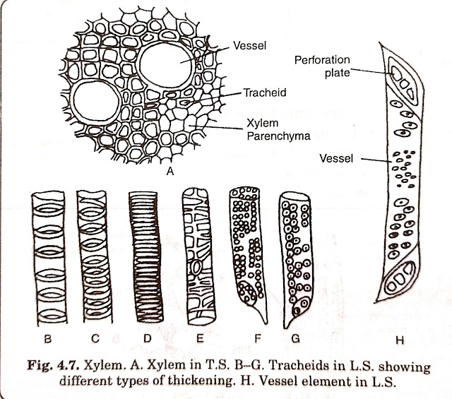
7. Xylem (Fig. 4.7)
Comments
(i) It is complex tissue, generally made up of three kinds of cells-xylem elements, xylem parenchyma and xylem fibres.
(ii) Xylem elements consists of two types of cells tracheids and vessels.
(iii) The tracheids are narrow elongated cells with annular, spiral, scalariform, reticulate and pitted thickening.Their end walls are tapering. The contact with the neighbouring tracheids is made through the common pit.
(iv) The vessels are also elongated and highly lignified. Their end walls have a simple perforation or multiple perforations. Their secondary walls possess pits.
(v) A few parenchyma cells and sclerenchyma fibers are also present in xylem.
8. Phloem (Fig. 4.8)
Comments
(i) It is a complex tissue made up of four types of cells-sieve elements, companion colls, phloem parenchyma and phloem fibres.
(ii) The sieve elements are elongated cells and are of two types-sieve cells and sieve tubes.
(iii) The sieve cells have sieve plates both on the side walls and on the end walls, while sieve tubes have sieve plates at the ends of the cells.
(iv) Adjacent to the sieve element, there lies an elongated companion cell with a prominent nucleus.
(v) A few parenchyma cells called phloem parenchyma and sclerenchyma fibres called phloem fibres are also present in the tissue.


0 Comments
Give me only suggestions and your opinion no at all Thanx