EXPERIMENT 5
Objective. To observe and make labelled diagrams of the microscopic structures of animal tissues (squamous epithelium, muscle fibres, nerve fibre and mammalian blood smear).
REQUIREMENTS
Microscope, permanent slides of cartilage, bone and muscle tissues, epithelial tissue and gonads.
OBSERVATIONS AND COMMENTS
1. Squamous Epithelium
Identification. W.M. (whole mount) of Squamous epithelium.
Comments
1.It consists of flat, plate like, polygonal cells.
2.The cells are fitted edge to edge without intercellular matrix.
3. Each cell has an oval or spherical nucleus in the centre that causes bulging of the cytoplasm.
4. Squamous epithelium forms the inner lining of lungs,alveoli and blood vessels, wall of Bowman's capsule's etc.

2. Striated or Skeletal Muscles (Voluntary Muscles)
Comments
1. The muscle fibres are elongated, cylindrical and lie parallel to each other.
2. Each fibre is multinucleated and is covered by a membrane called sarcolema.
3. Each fibre is filled with sarcoplasm, in which numerous myofibrils are arranged lengthwise.
4.Each myofibril is differentiated into light coloured I-band and dark coloured A-band, which produce cross striations in muscle fibres.
5.Striated muscles are attached to the bones and are present in the general musculature of the body. They are under the control of the will of animal.

3. Non-striated or Smooth Muscles (Involuntary muscles)
Comments
1.The muscle fibres are elongated, spindle shaped and are arranged into bundles.
2.Each muscle fibre has an oval shaped nucleus in the thickened middle part and is covered by a thin membrane called sarcolemma.
3.The sarcoplasm (cytoplasm) contains numerous thin myofibrils running lengthwise. Transverse striations are absent.
4.The relaxed fibres appear thin and elongated while the contracted ones appear thick and short.
5. Non-striated muscles occurs in the walls of alimentary canal, blood vessels, urinary and genital ducts and are not under the control of will.

4. Cardiac Muscles
Identification. W.M. of Cardiac muscles.
Comments
1.The muscle fibres are cylindrical branched and anastomosing.
2.Each fibre is covered with sarcolemma and may be uninucleate or binucleate.
3.The muscle fibres have longitudinal striations along with faint cross striations in the sarcoplasm.
4.The fibres are characterised by the presence of intercalated discs, where two fibres or their branches join.
5. Cardiac muscles are found in heart only.
6. The cardiac muscles are involuntary and work continuously throughout the life without getting tired.
5. Nerve (Neuron)
Identification. W.M. of Nerve cell (Neuron).
Comments
1.It consists of the cell body called cyton and the nerve fibre (axon).
2.There are present a prominent nucleus, Nissl's granules and neurofibrils in the cell body.
3.A number of processes called neurites arise from the cell body.
4.The neurites are of two types-shorter dendrites or dendrons and a long axon (neuraxon). The
fine branches of dendron may be one to several but axon is always one.
5. The axon ends in a group of branches called terminal arborizations (axon endings) certain axons
also give rise to side branches called collateral
fibres.
6.The axon part of a neuron may be covered by a medullary sheath or myelin sheath, which possess nodes of Ranvier.
7.The medullary sheath and nodes of Ranvier are surrounded by a transparent cellular covering
known as neurilemma of schwann cell.
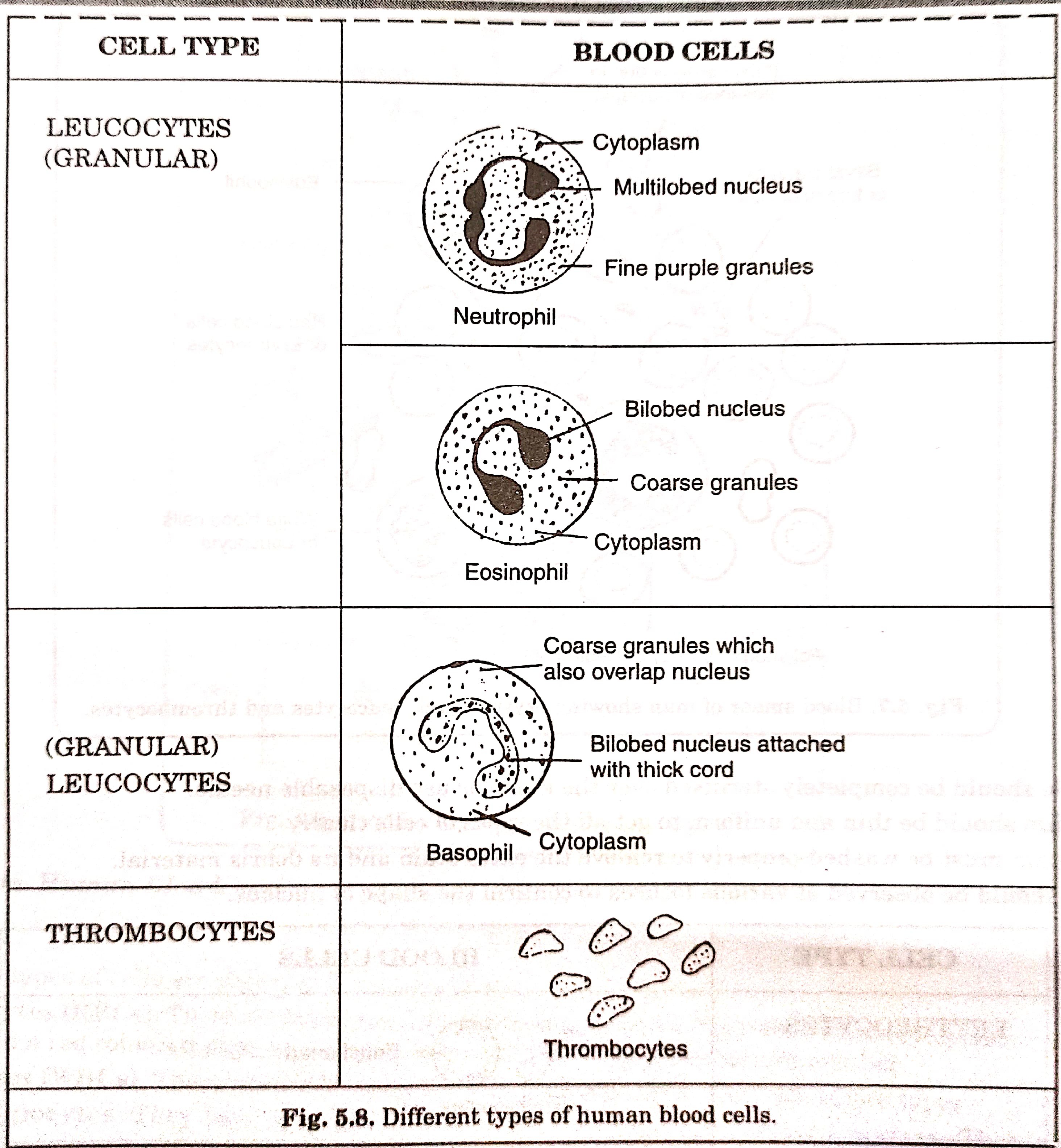
6. Identification. Human Blood.
Comments
Following types of cells are observed in human blood:
1.Erythrocytes (RBCs). These are biconcave, disc like enucleated cells. They are without cytoplasmic organelles but contain a red coloured pigment-haemoglobin. They are seen in maximum number.
2. Leucocytes (WBCs). These are large amoeboid and nucleated cells. They are of two types:
(a) Agranulocytes. They lack granules in their cytoplasm and have unlobed nucleus. They are of two types:
(i) Lymphocytes. They have spherical and prominent nucleus that fills most of the cytoplasm. They are
stained dark purple.
(ii) Monocytes. They are large cells with kidney shaped nucleus. They are also stained dark purple.
(b) Granulocytes. They have granules in their cytoplasm and possess lobed nucleus. They are of three types:
(i) Neutrophils. They are quite large cells with multilobed nucleus. They are stained orange red.
(ii) Eosinophils or Acidophils. They are also fairly large cells, but possess bilobed nucleus. They are
stained orange red.
(iii) Basophils. They are very less in number and possess S-shaped nucleus. They are stained dark blue.
Thrombocytes or Platelets. They are small ovoid or stellate, enucleated bodies. They appear as violet
granules.
PRECAUTIONS
1. Finger must be sterilised properly before pricking.
2. Needle should be completely sterilised over the flame or use disposable needle.
3.The film should be thin and uniform to get all the types of cells clearly.
4.The stain must be washed properly to remove the extra stain and it's debris material.
5.cells should be observed at various focuses to conferm the shape of nucleus.


.)
0 Comments
Give me only suggestions and your opinion no at all Thanx